

 menu
menu


raw data letters
 | IUCrDATA |
 access
Developed community standards on setting the diffraction resolution yields improved density maps and a new monosaccharide model in pdb_00004dda

aDepartment of Chemistry, University of Manchester, Manchester, M13 0PL, United Kingdom, bDivision of Biochemistry, Netherlands Cancer Institute, 1066CX Amsterdam, The Netherlands, and cUtrecht University, 3584 CG Utrecht, The Netherlands
*Correspondence e-mail: [email protected]
A systematic survey of in the PDB archive threw up a possible improvement in model fit in pdb_00004dda. Reprocessing of the publicly open archive of the raw diffraction images was made using EVAL. The diffraction resolution was assessed by the CC1/2 = 0.5 criterion and confirmed by paired model implemented in PDB-REDO. The resulting change in diffraction resolution, from 2.4 Å to 2.0 Å, improved the clarity of the electron density at the monosaccharide binding site. The NAG (N-acetyl-β-D-glucosamine) was refitted as NDG (N-acetyl-α-D-glucosamine). This example illustrates the advantages of raw diffraction data archiving, as aimed at by FAIR data polices, allowing reprocessing as agreement on community metrics.
Keywords: raw diffraction data archiving; community data metrics; raw data reprocessing; electron-density model fitting.
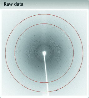
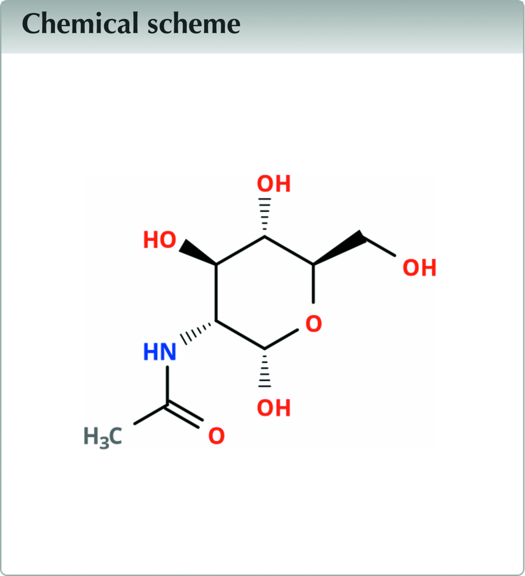
Metadata imgCIF file: https://doi.org/10.1107/S2414314626008515/ii4005img.cif
Introduction
A systematic survey of in the PDB archive threw up a possible improvement in model fit in pdb_00004dda. The structure was published in Tanley et al. (2012 ) with a diffraction resolution of <I/σI> = 4.0. The ways to establish the diffraction resolution have since then developed into community-agreed improved criteria. Reprocessing of the publicly open archive of the raw diffraction images was made using EVAL (Schreurs et al. 2010). The diffraction resolution was assessed by the CC1/2 = 0.5 criterion (Karplus & Diederichs 2012) and confirmed by paired-model refinement using Refmacat (Yamashita et al. 2023) implemented in PDB-REDO (Joosten et al. 2014). The resulting change in diffraction resolution, from 2.4 Å to 2.0 Å, improved the clarity of the electron density at the monosaccharide binding site. The NAG (N-acetyl-β-D-glucosamine) was refitted as NDG (N-acetyl-α-D-glucosamine). The raw diffraction images for pdb_00004dda in the original RAXIS.osc format, along with the reprocessing files, can be found at https://zenodo.org/records/18258544. The raw data were converted to fullCBF with the make-cbf tool as part of imgCIF_Creator (Kluyver et al., 2026) and deposited to Zenodo (https://zenodo.org/records/20326720). This example illustrates the advantages of raw diffraction data archiving, as aimed at by FAIR data polices (Wilkinson et al., 2016), allowing reprocessing as agreement on community metrics improve.
The benefits of raw diffraction data archiving linked to deposited PDB files were described with an example by the IUCr abstract (Helliwell et al., 2023) as a case study within the Global Open Science Cloud (Chen et al. 2023). Their example documented a case of a good choice of diffraction limit by the authors (Sato et al. 2021) and PDB entry pdb_00007ccy. In the case of pdb_00004dda we can now illustrate below the corrective power and an improvement with one of our own studies.
Methods and results
Data were processed in the same way as in Tanley et al. (2012) and detailed in Tanley et al. (2013) by EVAL (Schreurs et al. 2010), which uses a diffraction data integration method based on ab initio predicted profiles, but only extending the resolution and with the summary statistics now including CC1/2. The new diffraction resolution was chosen to be at a CC1/2 criterion of 0.5 and confirmed by paired model implemented in PDB-REDO (Joosten et al. 2014). The electron density at the monosaccharide binding site after the reprocessing and re-refinement is shown in Fig. 1. The raw data collection details are provided in Table 1. The data statistics of pdb_00004dda and of the reprocessed and re-refined models are given in Table 2. The metadata for the raw diffraction images have been automatically checked by the IUCrData checkcif-for-raw-data software (https://publbio.iucr.org/imgcif/).
| ||||||||||||||||||||||||||||||||||||||||||||||||
|
![[Figure 1]](ii4005fig1thm.gif) | Figure 1 Electron density 2mFo−DFc (1.58 r.m.s.) with the monosaccharide NDG fitted (this study at 2.0 Å). The of NDG is 0.93. This figure was produced using Coot (Emsley et al., 2010 ). |
Discussion and conclusions
As community metrics in macromolecular crystallography evolve, the processing of archived raw diffraction images can be successfully re-evaluated as shown in this case with an improved diffraction resolution limit and model. The full range of situations suitable for a Raw Data Letter are described in Kroon-Batenburg et al. (2022).
Supporting information
mtz file. DOI: https://doi.org/10.1107/S2414314626008515/ii4005sup1.txt
mmCIF file for structure 2WIY. DOI: https://doi.org/10.1107/S2414314626008515/ii4005sup2.txt
Metadata imgCIF file. DOI: https://doi.org/10.1107/S2414314626008515/ii4005img.cif
CheckCIF for raw data report. DOI: https://doi.org/10.1107/S2414314626008515/ii4005img_check.pdf
Acknowledgements
We thank Jasmine Young of the RCSB for assistance. Simon Tanley was supported by an EPSRC PhD studentship in the Department of Chemistry, University of Manchester. We thank James Hester for writing a tool to convert RAXIS.osc images to fullCBF.
Data availability
The raw diffraction data and the reprocessed data to 2.0 Å using EVAL as well as the molecular model are available at https://zenodo.org/records/18258544. The raw diffraction images converted to fullCBF format are available at https://zenodo.org/records/20326720. The improved Protein Data Bank entry can be found at pdb_000030IG. The pdb_00004dda entry is now obsolete.
References
 Chen, Y., Zhang, L., Li, J., Hodson, S., Uhlir, P. F, Li, X., Pergl, H., Haagstrom, I., Yue, X., Wang, J., Zhang, X., Helliwell, J. R., Kurisu, G., Miller, Yingchao Piao, Monthip Sriratana, Gensuo Jia, Bapon Fakhruddin, Zhang Ying, J., Bouwman, J., Liu, L., Maxwell, L., Oladipo, F., Sithole, H., Luo, Z. & Zhang, H. (2023). The Global Open Science Cloud: Vision and Initial Successes. GOSC IPO, 25 Aug 2023. https://doi.org/10.5281/zenodo.8296517. Google Scholar
Emsley, P., Lohkamp, B., Scott, W. G. & Cowtan, K. (2010). Acta Cryst. D66, 486–501. Web of Science CrossRef CAS IUCr Journals Google Scholar
Helliwell, J. R., Kurisu, G. & Kroon-Batenburg, L. (2023). Acta Cryst. A79, C822. CrossRef IUCr Journals Google Scholar
Joosten, R. P., Long, F., Murshudov, G. N. & Perrakis, A. (2014). IUCrJ 1, 213-220. Web of Science CrossRef PubMed IUCr Journals Google Scholar
Karplus, P. A. & Diederichs, K. (2012). Science 336, 1030–1033. Web of Science CrossRef CAS PubMed Google Scholar
Kluyver, T., Dall'Antonia, F., Hester, J. R. & Kroon-Batenburg, L. M. J. (2026). imgCIF_Creator. https://github.com/COMCIFS/imgCIF_Creator. Google Scholar
Kroon-Batenburg, L. M. J., Helliwell, J. R. & Hester, J. R. (2022). IUCrData 7, x220821. Google Scholar
Sato, H., Sugishima, M., Tsukaguchi, M., Masuko, T., Iijima, M., Takano, M., Omata, Y., Hirabayashi, K., Wada, K., Hisaeda, Y. & Yamamoto, K. (2021). Biochem. J. 478, 1023–1042. Web of Science CrossRef CAS PubMed Google Scholar
Schreurs, A. M. M., Xian, X. & Kroon-Batenburg, L. M. J. (2010). J. Appl. Cryst. 43, 70–82. Web of Science CrossRef CAS IUCr Journals Google Scholar
Tanley, S. W. M., Schreurs, A. M. M., Kroon-Batenburg, L. M. J., Meredith, J., Prendergast, R., Walsh, D., Bryant, P., Levy, C. & Helliwell, J. R. (2012). Acta Cryst. D68, 601–612. Web of Science CrossRef CAS IUCr Journals Google Scholar
Tanley, S. W. M., Schreurs, A. M. M., Helliwell, J. R. & Kroon-Batenburg, L. M. J. (2013). J. Appl. Cryst. 46, 108–119. Web of Science CrossRef CAS IUCr Journals Google Scholar
Wilkinson, M. D., Dumontier, M., Aalbersberg, I. J., Appleton, G., Axton, M., Baak, A., Blomberg, N., Boiten, J., da Silva Santos, L. B., Bourne, P. E., Bouwman, J., Brookes, A. J., Clark, T., Crosas, M., Dillo, I., Dumon, O., Edmunds, S., Evelo, C. T., Finkers, R., Gonzalez-Beltran, A., Gray, A. J. G., Groth, P., Goble, C., Grethe, J. S., Heringa, J., 't Hoen, P. A. C., Hooft, R., Kuhn, T., Kok, R., Kok, J., Lusher, S. J., Martone, M. E., Mons, A., Packer, A. L., Persson, B., Rocca-Serra, P., Roos, M., van Schaik, R., Sansone, S., Schultes, E., Sengstag, T., Slater, T., Strawn, G., Swertz, M. A., Thompson, M., van der Lei, J., van Mulligen, E., Velterop, J., Waagmeester, A., Wittenburg, P., Wolstencroft, K., Zhao, J. & Mons, B. (2016). Sci. Data3, 160018. Google Scholar
Yamashita, K., Wojdyr, M., Long, F., Nicholls, R. A. & Murshudov, G. N. (2023). Acta Cryst. D79, 368–373. Web of Science CrossRef IUCr Journals Google Scholar
This is an open-access article distributed under the terms of the Creative Commons Attribution (CC-BY) Licence, which permits unrestricted use, distribution, and reproduction in any medium, provided the original authors and source are cited.
| IUCrDATA |